Prenatal Heart Diagnosis: Methods & Testing Explained

Introduction
Did you know that prenatal heart diagnosis has advanced so dramatically that doctors can now detect up to 90% of major heart conditions before birth? As a pediatric cardiologist, I’ve witnessed how early detection transforms outcomes for families. Gone are the days of waiting until birth to identify cardiac concerns—modern imaging techniques offer an unprecedented view of your baby’s developing heart.
In this article, we’ll explore the methods and testing available in 2024 for prenatal heart diagnosis, helping you understand how these tools empower expectant parents and healthcare teams to plan for the best possible outcomes.
Early Screening Methods
Early screening methods play a pivotal role in identifying potential cardiac abnormalities during pregnancy. These methods are typically performed during the first and second trimesters to assess fetal development.
First-Trimester Screening
During the first trimester, a combination of ultrasound and maternal blood tests provides critical insights.
- Nuchal translucency ultrasound: Measures the fluid at the back of the baby’s neck, which may indicate chromosomal abnormalities linked to heart defects.
- Accuracy: While first-trimester screening can detect some signs of heart abnormalities, its overall accuracy improves when combined with genetic testing.
Routine Ultrasound in Cardiac Screening
Routine ultrasounds, particularly the anomaly scan conducted between 18-22 weeks, are essential for detecting structural abnormalities in the heart.
- Scope: These ultrasounds can identify four-chamber heart views and large vessel anomalies, providing a preliminary look at the fetal heart.
Timeline for Prenatal Heart Screenings
- First Trimester (11-14 weeks): Initial blood work and nuchal translucency ultrasound.
- Second Trimester (18-22 weeks): Detailed anomaly scan to assess structural cardiac issues.
- Third Trimester: Monitoring growth and function in cases of high-risk pregnancies.
Integration of Genetic Screening
Genetic screening methods, such as non-invasive prenatal testing (NIPT), assess chromosomal conditions that may correlate with congenital heart defects.
- Example: Trisomy 21 (Down syndrome) is linked to atrioventricular septal defects.
Advanced Diagnostic Imaging
For a more detailed evaluation, advanced imaging techniques provide high-resolution insights into fetal cardiac structure and function.
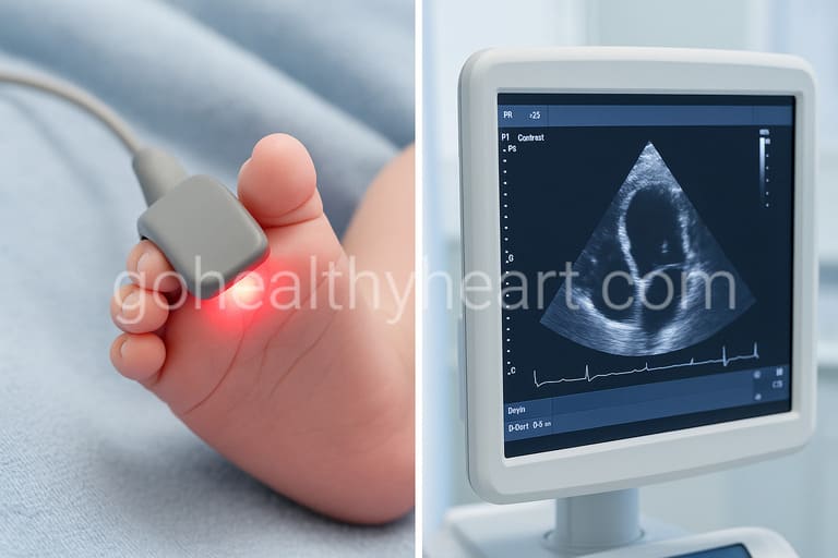
Fetal Echocardiography
Fetal echocardiography is the gold standard for prenatal heart diagnosis, offering a detailed view of the fetal heart.
- Timing: Typically performed between 18-24 weeks of pregnancy.
- Indications: Recommended for high-risk pregnancies or abnormal findings on routine ultrasound.
- Benefits: Detects conditions like ventricular septal defects, transposition of the great arteries, and hypoplastic left heart syndrome.
3D/4D Cardiac Ultrasound
Three-dimensional (3D) and four-dimensional (4D) ultrasounds enhance visualization of cardiac structures.
- Applications: Assess valve movements, ventricular function, and spatial orientation of vessels.
- Advantage: Real-time imaging helps in better understanding complex abnormalities.
Doppler Imaging
Specialized Doppler imaging evaluates blood flow within the fetal heart and great vessels.
- Purpose: Identifies issues like abnormal blood flow patterns, which can signal conditions like coarctation of the aorta.
Fetal Cardiac MRI
Although not as commonly used, fetal cardiac MRI provides additional insights in challenging cases.
- Advancements: High-resolution imaging of extracardiac structures such as the lungs and diaphragm.
- Use Case: Particularly helpful when echocardiographic images are suboptimal.
Risk-Based Testing Protocols
Certain pregnancies require enhanced monitoring based on maternal, familial, or environmental risk factors.
Maternal Conditions
Conditions like diabetes, lupus, or phenylketonuria can increase the risk of congenital heart defects.
- Example: Pre-gestational diabetes raises the likelihood of structural defects like tetralogy of Fallot.
Family History
A family history of congenital heart disease warrants specialized testing.
- Example: Siblings with congenital heart defects increase recurrence risks by 2-3 times.
Environmental and Medication-Related Risks
- Teratogenic medications: Certain drugs, like isotretinoin, are known to cause fetal cardiac defects.
- Environmental factors: Exposure to smoking or alcohol can also impact heart development.
Guidelines for High-Risk Pregnancies
High-risk pregnancies benefit from a tailored protocol, including more frequent ultrasounds and fetal echocardiograms.
Understanding Diagnostic Results
After diagnostic testing, interpreting results is a crucial step in planning care for the baby and mother.
Types of Cardiac Findings
- Structural abnormalities: Issues like septal defects or valve malformations.
- Functional concerns: Problems with the heart’s ability to pump or regulate blood flow.
Commonly Diagnosed Heart Conditions
Prenatal screening often identifies conditions such as:
- Hypoplastic left heart syndrome
- Tetralogy of Fallot
- Transposition of the great arteries
Accuracy Rates and Limitations
While modern imaging is highly accurate, certain limitations exist.
- False positives/negatives: In some cases, conditions may be missed or misinterpreted.
- Follow-up testing: Additional postnatal tests may be required for confirmation.
When Additional Testing is Needed
If results suggest a significant issue, your healthcare team may recommend advanced genetic testing or additional imaging closer to delivery.
Planning Your Care Journey
Once a diagnosis is made, a well-coordinated care plan ensures the best possible outcomes for both mother and baby.
Collaborative Care Teams
A multidisciplinary team of obstetricians, pediatric cardiologists, and maternal-fetal medicine specialists works together to monitor and manage the pregnancy.
Specialized Delivery Plans
For severe heart conditions, delivery planning may include:
- Timing adjustments: Earlier delivery to prevent complications.
- Specialized facilities: Delivery at a hospital equipped for immediate neonatal cardiac care.
Fetal Cardiac Interventions
In some cases, fetal cardiac interventions may be recommended.
- Examples: Balloon dilation for valve stenosis or in utero septostomy.
- Advances: Emerging techniques improve outcomes in select conditions.
Resources for Families
Navigating a prenatal heart diagnosis can be overwhelming, but resources like counseling, support groups, and specialized care centers can provide invaluable assistance.
Conclusion
Prenatal heart diagnosis has revolutionized the way we care for babies with congenital heart conditions. With early detection, families can prepare for the journey ahead, ensuring their baby receives the best care from the moment of birth. Advanced imaging techniques like fetal echocardiography and 3D/4D ultrasounds, combined with collaborative care, provide hope and clarity in challenging times.
Remember, if you have concerns about your baby’s heart health or are at increased risk, discuss advanced screening options with your healthcare provider. Early intervention is key to improving outcomes and giving your baby the healthiest start possible.
References: